Description
This project is an infographic explaining the segmentation clock, a complex concept in regenerative medicine, in a clear and engaging way. The layout draws inspiration from magazines like Scientific American, making the topic approachable for readers who are interested in science but not familiar with this area of research. Using visual storytelling, the fold-out design breaks down how the segmentation clock works and why it matters. It also connects current research to clinical trials as of February 2025, giving context for how these advances could influence future therapies.
Client
Shehryar Saharan (Prof. University of Toronto)
Year
2025
Audience
Scientists
Tools
Maya, Procreate, Illustrator
Type of Work
Coursework
Approach
Magazine spread with fold out
Research and Ideation
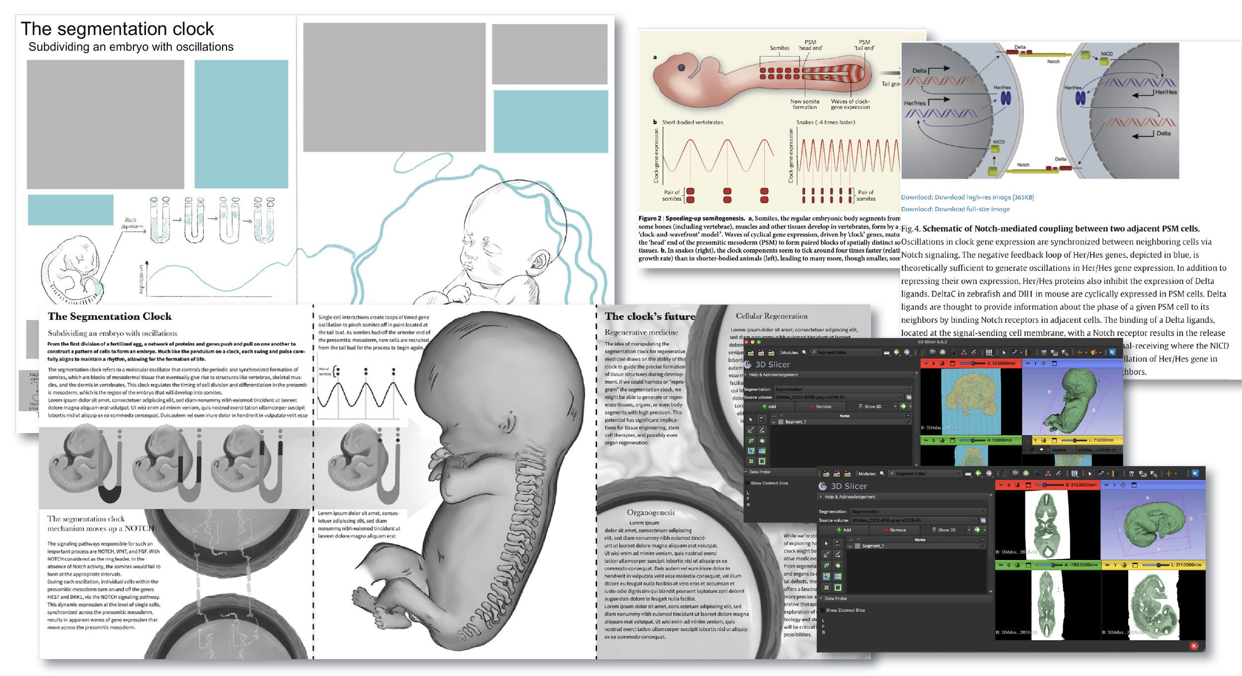
The research for this infographic was iterative, with the layout evolving alongside a deeper understanding of the segmentation clock. As I worked through the literature, key ideas and stages of the process were identified and prioritized, which shaped how the information was organized and presented visually. This back-and-forth between research and design helped the final layout reflect both the complexity of the topic and a clear narrative flow.
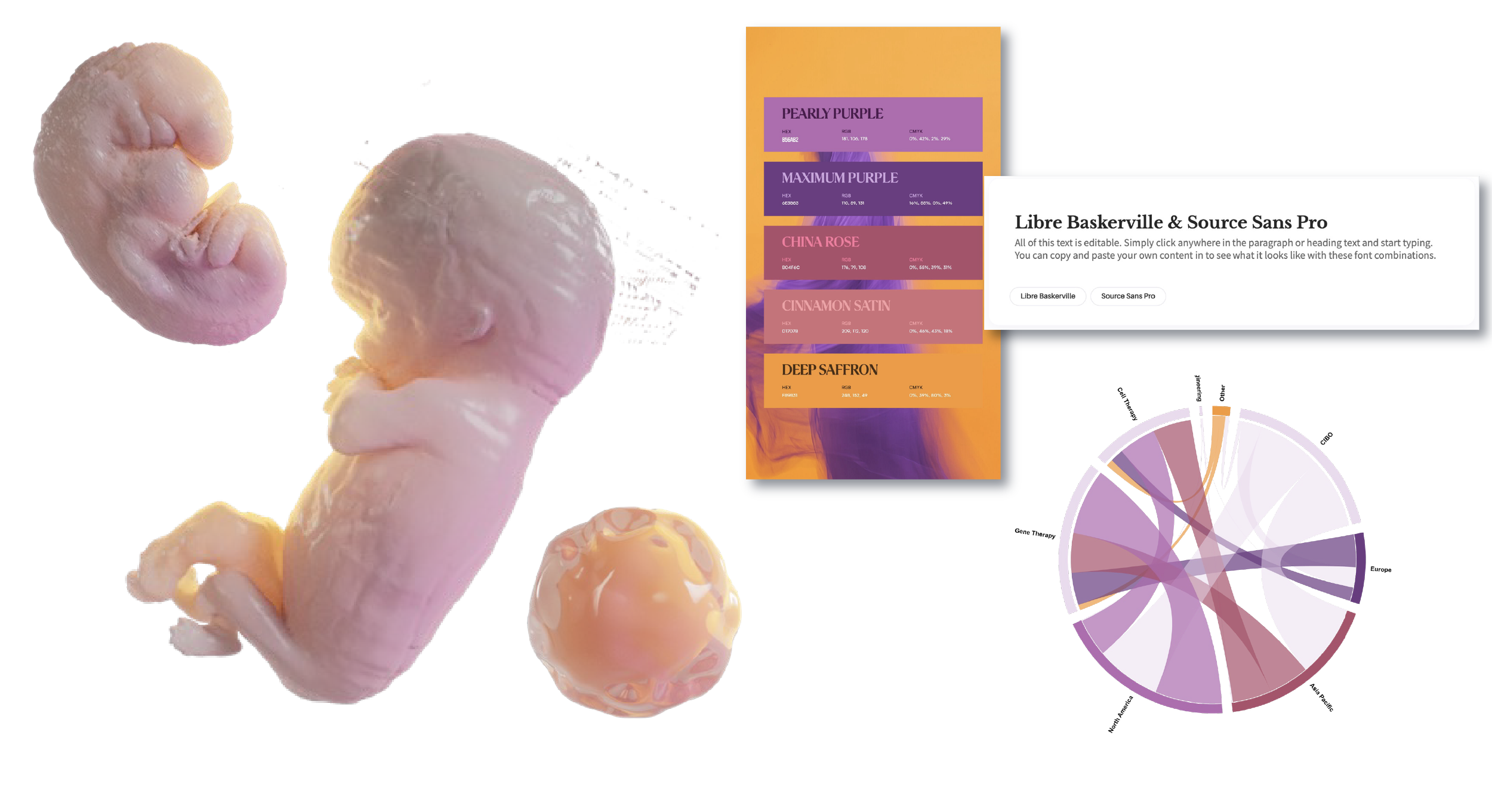
At the same time, I used 3D datasets to support anatomical accuracy. Files from the 3D Embryo Atlas were imported into 3D Slicer, where components were aligned and assembled to better understand spatial relationships during development. These models informed the visual breakdowns in the infographic, helping translate the science in a way that is both accurate and accessible.
Production
During production, the 3D models from 3D Slicer were brought into Autodesk Maya and refined through lighting, colour, and material adjustments to better define form, depth, and hierarchy. This also helped clean up noise and artifacts from the raw outputs, making the anatomy easier to read. These renders acted as a base rather than final images. After exporting, they were painted over digitally to simplify forms, improve clarity, and introduce a more cohesive, painterly style. This hybrid 3D-to-2D workflow kept the anatomy accurate while making the visuals clear and accessible.
References
Ajmal, L., Ajmal, S., Ajmal, M., Nawaz, G., Ajmal, L., Ajmal, S., Ajmal, M., & Nawaz, G. (2023). Organ Regeneration Through Stem Cells and Tissue Engineering. Cureus, 15(1). https://doi.org/10.7759/CUREUS.34336
Alliance for Regnerative Medicine. (2025). Clinical Trials by Therapeutic Approach - 2024 Q4.
Baldwin, C., Kim, J., Sivaraman, S., & Rao, R. R. (2022). Stem cell-based strategies for skeletal muscle tissue engineering. Journal of Tissue Engineering and Regenerative Medicine, 16(12), 1061–1068. https://doi.org/https://doi.org/10.1002/term.3355
Diaz-Cuadros, M., Wagner, D. E., Budjan, C., Hubaud, A., Tarazona, O. A., Donelly, S., Michaut, A., Al Tanoury, Z., Yoshioka-Kobayashi, K., Niino, Y., Kageyama, R., Miyawaki, A., Touboul, J., & Pourquié, O. (2020). In vitro characterization of the human segmentation clock. Nature, 580(7801), 113–118. https://doi.org/10.1038/s41586-019-1885-9
FontPair.co. (n.d.). Retrieved February 25, 2025, from https://www.fontpair.co/pairings/libre-baskerville-source-sans-pro
Gibb, S., Maroto, M., & Dale, J. K. (2010). The segmentation clock mechanism moves up a notch. Trends in Cell Biology, 20(10), 593–600. https://doi.org/10.1016/j.tcb.2010.07.001
Home | 3datlas. (n.d.). Retrieved February 4, 2025, from https://www.3dembryoatlas.com/
Infographics - Mesa Schumacher. (n.d.). Retrieved February 25, 2025, from https://www.mesaschumacher.com/infographic-projects/
Kwan, M. D., & Longaker, M. T. (2008). Regenerative Medicine: The Next Frontier. Transplantation, 86(2). https://journals.lww.com/transplantjournal/fulltext/2008/07270/regenerative_medicine__the_next_frontier.6.aspx
Maroto, M., Bone, R. A., & Dale, J. K. (2012). Somitogenesis. Development, 139(14), 2453–2456. https://doi.org/10.1242/dev.069310
Matsuda, M., Yamanaka, Y., Uemura, M., Osawa, M., Saito, M. K., Nagahashi, A., Nishio, M., Guo, L., Ikegawa, S., Sakurai, S., Kihara, S., Maurissen, T. L., Nakamura, M., Matsumoto, T., Yoshitomi, H., Ikeya, M., Kawakami, N., Yamamoto, T., Woltjen, K., … Alev, C. (2020). Recapitulating the human segmentation clock with pluripotent stem cells. Nature, 580(7801), 124–129. https://doi.org/10.1038/s41586-020-2144-9
RAWGraphs. (n.d.). Retrieved February 25, 2025, from https://www.rawgraphs.io
Search listening tool for market, customer & content research - AnswerThePublic. (n.d.). Retrieved February 1, 2025, from https://answerthepublic.com/
Shapira, A., & Dvir, T. (2021). 3D Tissue and Organ Printing—Hope and Reality. Advanced Science, 8(10), 2003751. https://doi.org/https://doi.org/10.1002/advs.202003751
Sonnen, K. F., Lauschke, V. M., Uraji, J., Falk, H. J., Petersen, Y., Funk, M. C., Beaupeux, M., François, P., Merten, C. A., & Aulehla, A. (2018). Modulation of Phase Shift between Wnt and Notch Signaling Oscillations Controls Mesoderm Segmentation. Cell, 172(5), 1079-1090.e12. https://doi.org/https://doi.org/10.1016/j.cell.2018.01.026
Venzin, O. F., & Oates, A. C. (2020). What are you synching about? Emerging complexity of Notch signaling in the segmentation clock. Developmental Biology, 460(1), 40–54. https://doi.org/https://doi.org/10.1016/j.ydbio.2019.06.024
Vonk, F., & Richardson, M. (2008). Serpent clocks tick faster. Nature, 454, 282–283. https://doi.org/10.1038/454282a
Yen, B. L., Hsieh, C.-C., Hsu, P.-J., Chang, C.-C., Wang, L.-T., & Yen, M.-L. (2023). Three-Dimensional Spheroid Culture of Human Mesenchymal Stem Cells: Offering Therapeutic Advantages and In Vitro Glimpses of the In Vivo State. Stem Cells Translational Medicine, 12(5), 235–244. https://doi.org/10.1093/stcltm/szad011