Description
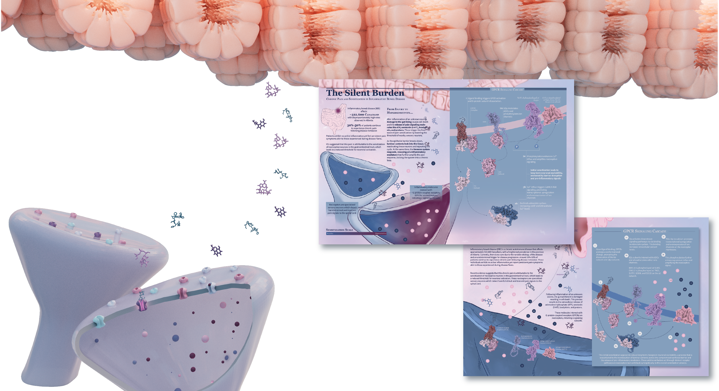
This two-page spread uses a molecular visualization to explore how pain becomes sensitized in inflammatory bowel disease (IBD). It focuses on how molecules released during cell damage interact with nociceptors, showing how chronic inflammation leads to an amplified pain response. The piece also highlights the cyclical nature of IBD pain, where ongoing inflammation activates nerves that, in turn, further drive inflammation. By visualizing this feedback loop, the spread helps explain why breaking the cycle is key to developing more effective pain-relief therapies.
Exhibited in:
AMI Annual Salon 2026
Tools
ChimeraX, VMD, Z-Brush, Maya, and Illustrator
Type of Work
Assignment
Approach
2 page magazine spread
Client
Dr Derek Ng (University of Toronto)
Year
2025
Audience
General
Research and Ideation
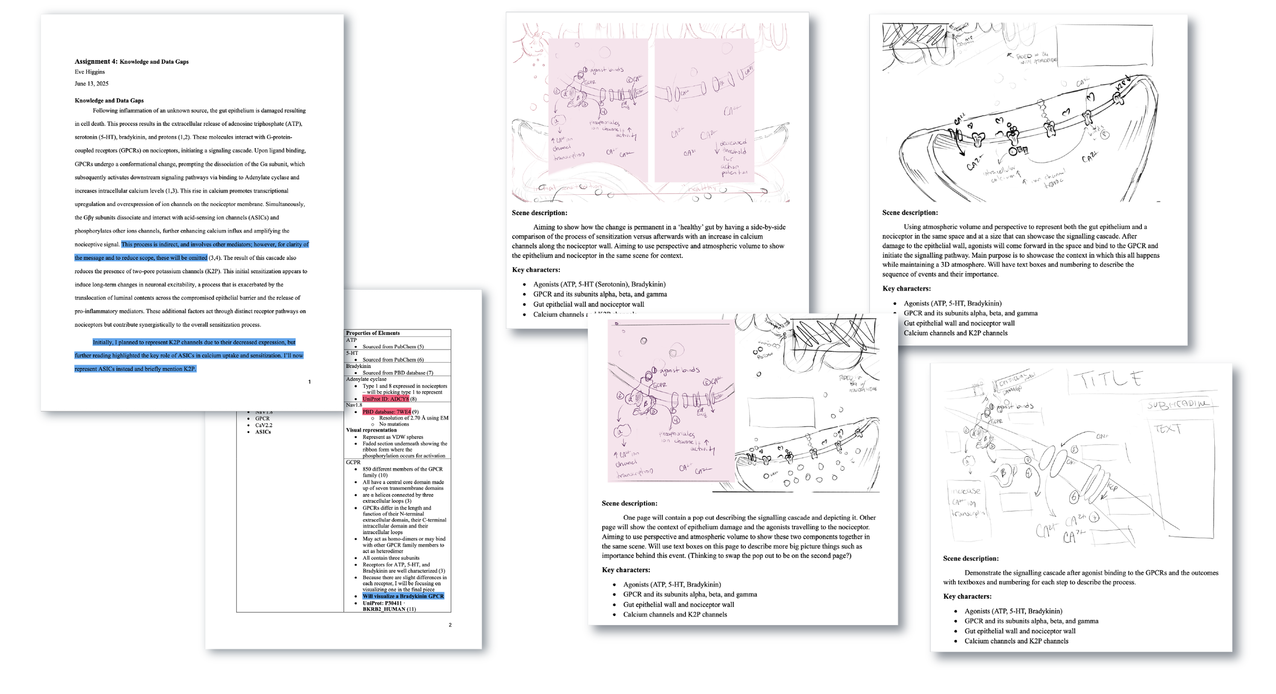
The development of this spread began with synthesizing current research on inflammatory bowel disease, with a particular focus on the molecular mechanisms underlying chronic pain sensitization. Literature reviews and discussions with subject matter experts informed which biological processes were most essential to communicate, guiding the selection of receptors, ion channels, and signalling pathways that would accurately represent nociceptor activation while remaining accessible to the intended audience.
Research and visual development evolved together through an iterative design process. As the scientific narrative became more defined, the layout was continually refined to establish a clear information hierarchy, balance visual complexity, and guide the reader through multiple layers of molecular interactions. Every design decision was made to transform dense scientific content into a cohesive visual story, one that preserves scientific accuracy while making complex mechanisms easier to interpret and remember.
Production
The production process combined molecular visualization, 3D modeling, and illustration to communicate complex biological interactions with both precision and clarity. Molecular structures were first prepared from structural datasets before being integrated into a three-dimensional environment where scale, spatial relationships, and composition could be carefully developed. Additional biological elements, including neurons, epithelial cells, and ion channels, were sculpted to create a cohesive visual ecosystem that accurately represented the underlying biology while supporting the overall narrative.
The final composition was assembled and refined through illustration, where visual hierarchy, colour, and composition were used to distinguish key signalling events and guide the viewer through the mechanism. By combining structurally accurate molecular data with custom-built anatomical models and thoughtful information design, the final spread transforms an intricate cellular process into a clear, engaging visual explanation without compromising scientific integrity.
References
Background information, Page 1:
Impact of IBD in Canada Report - Impact of IBD in Canada Report - Crohn’s and Colitis Canada. Available from: https://crohnsandcolitis.ca/About-Us/Resources-Publications/Impact-of-IBD-Report
Szigethy E. Pain Management in Patients With Inflammatory Bowel Disease. Gastroenterol Hepatol (N Y). 2018 Jan;14(1):53. Available from: https://pmc.ncbi.nlm.nih.gov/articles/PMC5824598/
Brierley SM, Linden DR. Neuroplasticity and dysfunction after gastrointestinal inflammation. Nature Reviews Gastroenterology & Hepatology 2014 11:10. 2014 Jul 8;11(10):611–27. Available from: https://www.nature.com/articles/nrgastro.2014.103
Grundy L, Erickson A, Brierley SM. Visceral Pain. Annu Rev Physiol. 2019 Feb 10;81(Volume 81, 2019):261–84. Available from: https://www.annualreviews.org/content/journals/10.1146/annurev-physiol-020518-114525
Signaling Cascade, Page 2:
Brierley SM, Linden DR. Neuroplasticity and dysfunction after gastrointestinal inflammation. Nature Reviews Gastroenterology & Hepatology 2014 11:10. 2014 Jul 8;11(10):611–27. Available from: https://www.nature.com/articles/nrgastro.2014.103
Molliver DC. G-Protein Coupled Receptors in Sensory Neuron Function and Pain. Encyclopedia of Neuroscience. 2009;1761–5. Available from: https://link.springer.com/rwe/10.1007/978-3-540-29678-2_2070
Gribkoff VK. The role of voltage-gated calcium channels in pain and nociception. Semin Cell Dev Biol. 2006 Oct 1;17(5):555–64. Available from: https://www.sciencedirect.com/science/article/abs/pii/S1084952106000991
Small Molecules Used:
ATP, sourced from PDB Database
RCSB PDB - ATP Ligand Summary Page. Available from: https://www.rcsb.org/ligand/ATP
5-HT, sourced from PubChem Serotonin | C10H12N2O | CID 5202 - PubChem. Available from: https://pubchem.ncbi.nlm.nih.gov/compound/5202
Bradykinin, sourced from PDB database: ID 7F6H RCSB PDB - 7F6H: Cryo-EM structure of human bradykinin receptor BK2R in complex
Gq proteins and bradykinin. Available from: https://www.rcsb.org/structure/7F6H
Proteins Used:
Adenylate cyclase, sourced from UniProt: ID ADCY8. Currently trying to find how it binds to the Gα subunit.
ADCY8 - Adenylate cyclase type 8 - Homo sapiens (Human) | UniProtKB | UniProt. Available from: https://www.uniprot.org/uniprotkb/P40145/entry#structure
Nomura, R., Suzuki, S., Nishikawa, K., Suzuki, H., & Fujiyoshi, Y. (2025). Structural insights into human adenylyl cyclase 9 in complex with Gαs by Cryo-EM. Journal of Structural Biology, 217(3), 108223. https://doi.org/10.1016/j.jsb.2025.108223 RCSB PDB - 9U3R: Cryo-EM structure of human AC9. Available from: https://www.rcsb.org/structure/9U3R
Nav1.8, sourced from PBD database: ID 7WE4 and its phosphorylation sites for activation.
RCSB PDB - 7WE4: Human Nav1.8 with A-803467, class I. Available from: https://www.rcsb.org/structure/7WE4
Wu DF, Chandra D, McMahon T, Wang D, Dadgar J, Kharazia VN, et al. PKCε phosphorylation of the sodium channel NaV1.8 increases channel function and produces mechanical hyperalgesia in mice. J Clin Invest. 2012 Apr 2;122(4):1306–15. Available from: http://www.jci.org
Heinle JW, Dalessio S, Janicki P, Ouyang A, Vrana KE, Ruiz-Velasco V, et al. Insights into the voltage-gated sodium channel, NaV1.8, and its role in visceral pain perception. Front Pharmacol. 2024 May 23;15:1398409.
Bradykinin binding GPCR, sourced from PBD database: ID 7F6H.
RCSB PDB - 7MIY: Human N-type voltage-gated calcium channel Cav2.2 at 3.1 Angstrom resolution. Available from: https://www.rcsb.org/structure/7MIY Shen J, Zhang D, Fu Y, Chen A, Yang X, Zhang H. Cryo-EM structures of human bradykinin receptor-Gq proteins complexes. Nat Commun. 2022 Dec 1;13(1).
Geppetti P, Veldhuis NA, Lieu TM, Bunnett NW. G Protein-Coupled Receptors: Dynamic Machines for Signaling Pain and Itch. Neuron. 2015 Nov 18; 88(4):635–49. Available from: https://www.sciencedirect.com/science/article/pii/S0896627315009800#fig5
CaV2.2, sourced from PBD Database: ID 7MIY and its phosphorylation sites for activation.
RCSB PDB - 7MIY: Human N-type voltage-gated calcium channel Cav2.2 at 3.1 Angstrom resolution. Available from: https://www.rcsb.org/structure/7MIY
Lacinova L, Mallmann RT, Jurkovičová-Tarabová B, Klugbauer N. Modulation of voltage-gated CaV2.2 Ca2+ channels by newly identified interaction partners. Channels. 2020 Jan 1;14(1):380. Available from: https://pmc.ncbi.nlm.nih.gov/articles/PMC7567506/